So Mr Blakemore - exactly what have you done for us?
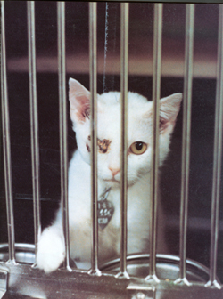
Colin Blakemore became Chief Executive of the MRC (Medical Research Council) on the 1st October 2003. He had previously been a Waynflete Professor of Physiology at Oxford University since 1979, which is where he became notorious for sewing up the eyes of kittens.

Colin Blakemore has managed throughout the years to capitalise on his high media profile and, despite his years of experiments having yielded nothing meaningful for the human problem associated with amblyopia and strabismus, he has become a media personality. Recent years have seen him with his own television series about the brain and a week seldom passes by without an appearance from Mr Blakemore on either television or radio. It's surprising what a careful manipulation of the media can do for ones celebrity status.

So what has Mr Blakemore actually done to enhance the font of medical knowledge? Mr Blakemore was born in 1944 in Stratford-upon-Avon and, if he is to be believed, and it's something he never seems to tire of telling us, into a Labour-supporting working class family. From such humble origins he has turned into a figure of such status that allowed him last year to publicly lambast the government for not having given him a knighthood. Not of course because he has any egotistical tendencies but because he deserves it! So you must now be asking yourself how many thousands of people has he managed to help with their eyesight problems in order that he can exert such a power over both the media and government. The following article debunks many of the falsehoods surrounding many of Mr Blakemore's more public pronouncements. We give you the facts and we let you decide the truth.
Once again SPEAK would like to take this opportunity to offer Mr Blakemore the chance to publicly put the record straight, an open and public debate into vivisection. It will give him a wonderful opportunity to tell the public exactly how vivisection has benefited them and just as importantly, exactly what he has done to benefit us all?
AMBLYOPIA: The failure of experiments on cats and monkeys
Blakemore's research has been into amblyopia and strabismus. Amblyopia is decrease in function of one or both eyes, or impairment in the way in which the eyes interact, for which no cause can be detected in the eyes. Strabismus is misalignment of the eyes, and can take the form of a squint.
Treatments and how they were discovered
Blakemore has claimed that advances have been due to research such as his: "...before the animal research began it was not known whether squint could cause amblyopia or whether the existence of poor vision in one eye causes the squint." [1] He also claims that amblyopia"...was poorly understood until about 25 years ago when experimental research involving animals began to tackle the question of its origin...in the absence of any real knowledge of the cause of the disease there was no single agreed method of treatment." [2]
In response to the first point, it should be noted that in the 1800s Claud Worth identified that is was the deficiency in eye function which caused the misalignment, or squint.[3] He was preceded by George Louis Leclerc Comte de Buffon, who did his research in the 1700s. [4]
Regarding treatment of amblyopia, this has changed little over recent years, and has not benefited from Blakemore's improved knowledge of blindness in kittens. The recognised pioneer in amblyopia treatment is Claude Worth, whose work is described thus: "It is surprising how little...has been added to the major store of knowledge as he presented it, at least from the practical viewpoint...Worth's emphasis on early treatment of the error of refraction and the amblyopia is still the keynote of the most rational approach to the treatment of the strabismic child..."[5] Worth retired fifty years before Blakemore started his experiments.
Worth's brilliant investigations include six essential points, still relevant, one of which is how amblyopia is acquired. He was aware of the danger of accidentally causing amblyopia of the better eye by treating children below the age of six.
Species differences
Research conducted on animals has been hampered by biological differences, which are insurmountable. Animals used have mainly been cats and monkeys, but chickens and tree shrews also have been used. Techniques have mainly been to sew the eyelids closed, remove an eye, cause artificial strabismus (either surgically or using prisms), or turn the material of the eye opaque.
The human eye depends on the fovea and macula, which cats do not have. These two areas are crucial in the study of amblyopia.[6] Researcher von Noorden commented "Human amblyopia involves primarily foveal vision rather than peripheral vision...".[7]
The neuroanatomy of the cat and human are different, highlighted by the lack of references in clinical research literature to neuroanatomical research in the cat.[8] The cat model has also proved to be unpredictive of human conditions.
The primate eye has been found to be of different composition from the human eye, as highlighted by Raviola and Wiesel. Their photographs have revealed the poor correlation of experimental monkeys in short-sightedness experiments with human myopia.[9] Animal experimenter Jampolsky has also noted that common forms of strabismus in humans are rarely, if ever, found naturally in monkeys. Aspects of the human condition include exotropia, accommodative esotropia, A and V patterns, dissociated vertical deviation and latent nystagmus. These are not features of induced and occurring money strabismus. [10]The human visual system is unique when studied at the detail necessary for knowledge to advance.
There's also differentiation between monkey species, as has been found by the experiments. Monkeys eyelids were sewn closed, to find that one species had eye elongation prevented by being given atropine, another did not.[11]
Inducing the fake condition
Problems are also caused by the way amblyopia is induced. In humans born with amblyopia, there are often complications caused by other abnormalities, which doesn't apply to animals. The animal work revolves around taking recordings from single cells, yet in humans vision has a high psychological element - a problem animal experimenters have admitted.[12] Cell recordings don't tell us what animals can see, or whether they see at all.
Sewing the eyelid shut differs from human blindness such as that caused by cataracts in the amount and quality of light reaching the retina. The human condition, which is present from birth, differs from the experimental condition, which is induced later. Primate experimenters have concluded that this artificial way of inducing the condition is a cause of differing results.[13] As all such methods are artificial, it is hard to see how faith in the animal method can be maintained.
Misleading
Animal studies have yielded results which have been found to be incorrect in application to humans. As one expert says "There has been considerable concern expressed by vision scientists other than just myself that the experimental work is either not relevant or may be misleading with respect to the human condition and its treatment."[14] Primate experiments have misled us over the critical time at which amblyopia develops.[15] A researcher expressed his hesitance to accept animal results, concluding "the morphologic and functional organization of the visual system in cats is substantially different from that in man." [16]
Boothe's team found two monkeys with congenital esotropia (eyes turned in), and discovered they had absent or small accessory lateral rectus (ALR) muscles.[17] Their conclusion that this was the cause of human esotropia has proved incorrect: patients are been found to have normal ALR muscles.
Monkey experiments suggested that children wearing eye patches might be harmful. Studies on children showed this was not the case. A researcher involved described this as a "surprise" and suggested that we should ask whether to believe animal results.[18]
A similar conclusion from cats prompted the response: "I...would hope that their results, based on the experimental technique described, were not interpreted to suggest that patching may do more harm than good, and would not influence clinicians to be less vigorous with their patching therapy. ...I think work will be necessary to more closely simulate the human clinical situation before further conclusions can be reached."[19]
Instead of vivisection
The animal method is not viable, but others are. Considering that all the results taken from animal experiments have so far been wrong, or already known, any future discoveries would have to be validated by the study of human patients - the method by which all advances have so far been made.
Technology now enables us to gain information by using non-invasive imaging technologies on patients. For example, modern magnetic resonance imaging technologies can assess visual cortex function with a resolution of 1.4 mm. [20] CAT scans, PET scans, and autopsy studies enable a more complete picture to be built up. The findings will be based on human neurology, as opposed to the very different neurology of an animal.
Resources available to study these proven and emerging, reliable techniques will be increased once the unscientific pre-occupation with animal experiments is abandoned.
References
- Colin Blakemore "A reply to criticism of experiments involving visual deprivation," September 1987.
- Colin Blakemore "A reply to criticism of experiments involving visual deprivation," September 1987.
- Abraham, S.V.: A tribute to Claud Worth. Ann. Ophthalmol. 4: 171-175,
1972.
- Marg, E.: Prentice Memorial Lecture: Is the animal model for stimulus deprivation amblyopia in children valid or useful? Am. J. Optometry Physiol. Optics 59: 451-464, 1982.
- Abraham, S.V.: A tribute to Claud Worth. An n. Ophthalmol. 4: 171-175, 1972.
- von Noorden, G .K.: Application of basic research data to clinical amblyopia. Ophthalmology 85: 496-504, 1978.
- von Noorden, G .K.: Application of basic research data to clinical amblyopia. Ophthalmology 85: 496-504, 1978.
- "AMBLYOPIA" Nedim C. Buyukmihci, V.M.D.a, http://avar.org/
- Raviola E, Wiesel TN. An animal model of myopia. New England Journal of Medicine 1985;312: 1609-1615.
- Jampolsky A. Unequal visual inputs and strabismus management: a comparison of human and animal strabismus, in Transactions of the New Orleans Academy of Ophthalmology, Symposium on Strabismus. St. Louis, CV Mosby, 1978, p 358-492
- Robinson CJ. Somatopographic organization in the second somatosensory area of M. fascicularis. Journal of Neumphysiology 1980; 192:43-67.
- Blakemore, C . and Vital-Durance, F.: Development of the neural basis of visual acuity in monkeys: Speculation on the origin of deprivation amblyopia. Trans. Ophthalmol. Soc. U.K. 99: 363-368, 1979.
- O'Dell CD, Gammon JA, Fernandes A, Wilson JR, Booth RG. Development of acuity in a primate model of human infant unilateral aphakia. Investigative Ophthalmology and Visual Science l989;30:2068-2074. Wilson JR, Tigges M, Booth RG, Tigges J, Gammon JA. Effects of aphakia on the geniculostriate system of infant rhesus monkeys. Acta Anatomica 1991; 142:193-203.
- "AMBLYOPIA" Nedim C. Buyukmihci, V.M.D.a, http://avar.org/
- Vaegan TD. Critical period for deprivation amblyopia in children. Transaction of the Ophthalmological Society of the United Kingdom 1979;99:432-439
- von Noorden, G .K. and Maumenee, A .E.: Clinic al observations on stimulus- deprivation amblyopia (amblyopia ex anopsia). Am. J. Ophthalmol. 65: 220-224, 1968.
- Boothe RG, Quick MW, Joosse MV, Abbas MA, Anderson DC. Accessory lateral rectus orbital geometry in normal and naturally strabismic monkeys. Investigative Ophthalmology and Visual Science 1990;31:1168-1174.
- Marg, E.: Prentice Memorial Lecture: Is the animal model for stimulus deprivation amblyopia in children valid or useful? Am. J. Optometry Physiol. Optics 59: 451-464, 1982. Hoyt, C.S .: The long- term visual effects of short-term binocular occlusion of at-risk neonates. Arch . Ophthalmol. 98: 1967-1970, 1980.
- Metz, Henry S.: To the editor. Invest. Ophthalmol. Vis . Sci. 26: 249, 1985
- Engel SE, Rumelhart DE, Wandell BA, et al. fMRI of human visual cortex. Nature 1994:369;525,
- Marg, E.: Prentice Memorial Lecture: Is the animal model for stimulus deprivation amblyopia in children valid or useful? Am. J. Optometry Physiol. Optics 59: 451-464, 1982.